SKIN CANCERS
Melanoma in situ
Confined to the top layers of skin, this type of melanoma has a cure rate approaching 100%.
- The most commonly diagnosed form of melanoma
- Spread to other parts of the body is rare
- Often detected at a routine skin check
- Treated with surgical removal

What is melanoma in situ?
Melanoma is an uncontrolled growth of melanocytes, the pigment-producing cells in the skin. If untreated, melanoma can grow deeper into the skin and spread to other parts of the body.
Melanoma in situ is melanoma at its earliest stage. The abnormal melanocytes are confined to the epidermis, the outer layer of the skin, and have not yet invaded deeper tissues.
The diagnosis of melanoma in situ is confirmed when a skin pathologist examines the removed lesion under a microscope and determines that the abnormal cells remain confined to the surface layer of the skin.
Prognosis of melanoma in situ
Melanoma outcome is strongly related to the thickness of the melanoma at the time of diagnosis.
Because melanoma in situ is confined to the outer layer of the skin, the prognosis is excellent and five-year survival approaches 100% when treated appropriately.
If left untreated, melanoma in situ can eventually grow deeper into the skin and develop into invasive melanoma, which has the potential to spread through blood vessels or lymphatic channels to other parts of the body.
Diagnosis of melanoma in situ
Approximately half of all melanomas are detected by the patient, who brings it to the attention of a skin cancer doctor. Usually, a melanoma in situ is flat, irregularly-shaped and dark. It may have multiple colours. It is often noticed because it looks different from a person’s other moles and spots, or because it is new, growing or otherwise changing.

MBBS, MA (Virtual Comm), Grad Cert Hlth Info, Grad Dip Comp Inf Sci
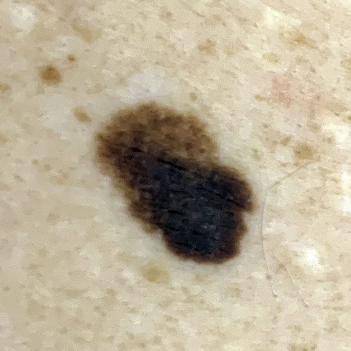
Close-up (non-dermoscopic) view of melanoma in situ
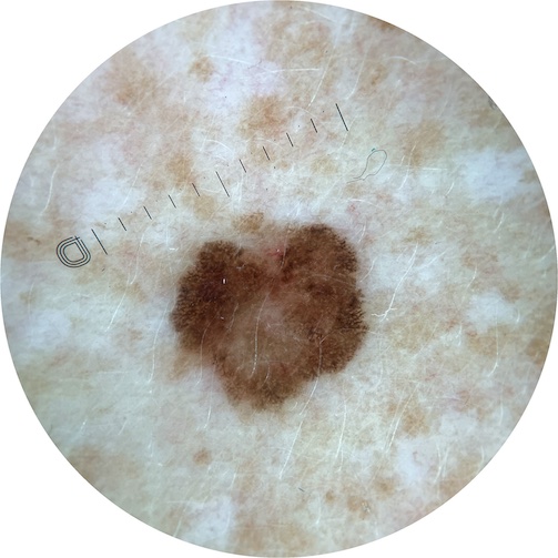
Dermoscopic view of melanoma in situ
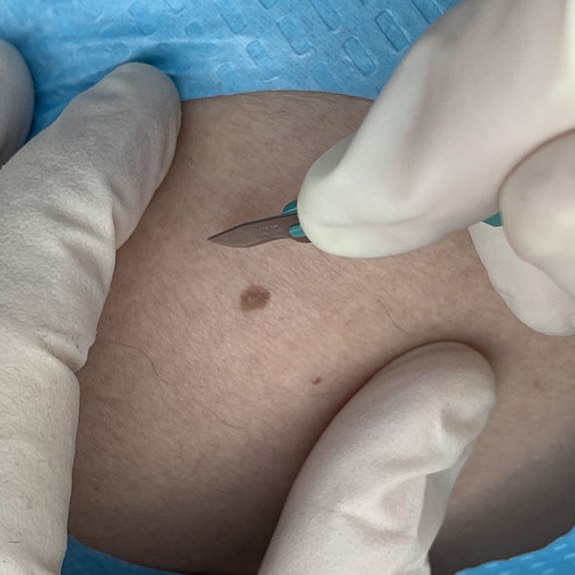
A skin cancer doctor can diagnose melanoma by examining it closely with a dermoscope. It will usually have specific colours or structures that alert the doctor to the possibility of melanoma. Sometimes these clues to diagnosis are hard to see and the melanoma is suspected because of changed appearance in repeated dermoscopic photographs. When a melanoma is suspected, the doctor will surgically remove the suspicious spot (a procedure known as excision biopsy) so that a skin pathologist can examine the tissue and make a definitive diagnosis.
Treatment of melanoma
All melanomas must be surgically removed with a clear margin around the edge. In most cases, melanoma in situ must be removed with a clear margin of at least 5 mm of normal skin around its outer edge. The initial excision biopsy is rarely this wide, so usually once a melanoma in situ is detected, a further excision must be performed to ensure a clearance of at least 5 mm. If the melanoma is in a cosmetically sensitive area or if the function or movement of the area is likely to be affected, your skin cancer doctor may refer you to a plastic surgeon or the melanoma unit of a hospital for further treatment.
Learn more about excision and excision biopsy.
Follow-up: what happens next?
Further assessment
The risk of a melanoma in situ spreading to other parts of the body is extremely low. Your doctor will perform a full skin examination to check for additional melanomas or other skin cancers elsewhere on the body.
Specialist referral
In some circumstances, your doctor may refer you to another doctor or health service for further treatment or assessment. For example:
- A plastic surgeon can perform a wider excision of the melanoma to achieve a good cosmetic outcome or preserve function of the affected area
- A public hospital melanoma unit can offer a team approach where surgeons, dermatologists, skin pathologists and other specialties are involved in your care. Your doctor may refer you to a public hospital melanoma unit for further treatment, to get a second opinion on the initial skin pathologist’s report, or advice on future follow-up and treatment.
Further tests
For melanoma in situ, routine blood tests or imaging scans are not usually required, as the risk of spread is extremely low.
Recurrence or spread after appropriately treated melanoma in situ is extremely unlikely. People who have had melanoma have an increased lifelong risk of a new melanoma.
High risk patients
Some people have an especially high risk of developing further melanomas in future. These people include:
- People who have already had more than one melanoma.
- People with a strong family history of melanoma.
Mole mapping (total body photography) is sometimes recommended for people in these high risk groups.
Self-examination
Skin cancer self-examination is an important part of ongoing care. People can detect new or changing lesions between appointments; report these promptly.
People with a history of melanoma should ideally check their own skin every 1–2 months.
Skin checks for your family
Melanoma tends to run in families. If you have had a melanoma, your first-degree relatives (children, sisters, brothers and parents) have an above-average chance of developing melanoma. They should check their own skin regularly and consider yearly skin checks by a doctor or nurse.
Further information
For detailed information about melanoma diagnosis, treatment and follow-up, see the Melanoma Institute Australia guide to melanoma in situ (PDF)
Skin checks in future
Follow-up looks for new melanomas and checks the treated site. Recurrence or spread after appropriately treated melanoma in situ is extremely unlikely; site and lymph-node examination are simple precautions during a general skin check.
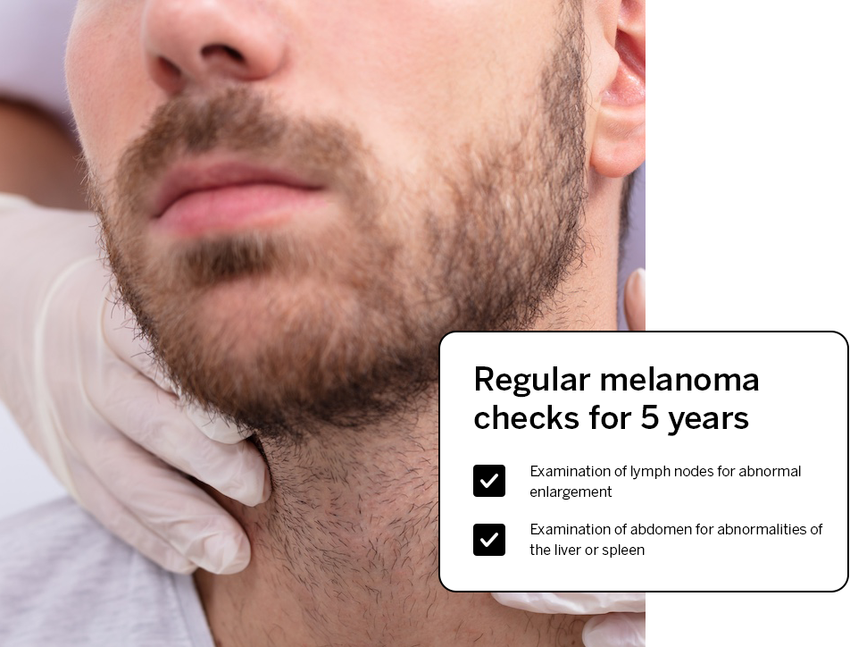
Every six months for two years: full body skin check, close inspection of the melanoma site and examination of nearby lymph nodes.
Yearly full body skin checks, continuing indefinitely. Your doctor may recommend closer review according to your individual risk.
Ongoing yearly skin checks are recommended for life.
References
Australian Institute of Health and Welfare. Skin cancer in Australia, 2016.
Menzies S, Guitera P, Soyer P et al. What is the role of sequential digital dermoscopy imaging in melanoma diagnosis? Clinical practice guidelines for the diagnosis and management of melanoma, 2018.
Sladden M, Nieweg O, Howle J et al. What are the recommended safety margins for radical excision of a primary melanoma (in situ)? Clinical practice guidelines for the diagnosis and management of melanoma, 2020.
Sladden M, Nieweg O, Howle J et al. Updated evidence-based clinical practice guidelines for the diagnosis and management of melanoma: definitive excision margins for primary cutaneous melanoma. Medical Journal of Australia volume208 issue3, February 2018, pages 137-142, 2018.
Barbour A, Guminski A, Liu W et al. Ideal settings, duration and frequency of follow-up for patients with melanoma. Clinical practice guidelines for the diagnosis and management of melanoma, 2019.
Concerned about a new or changing spot?
Dermoscopic examination and digital photography help detect melanoma at an early stage, when treatment is usually straightforward and highly successful.
Arrange a 1–3 spot assessment