LEARN
Dermoscopy and dermoscopic photography: detecting skin cancer clues
A dermoscopic photograph is a close-up image of a skin lesion taken with a dermoscope, usually using polarised light.
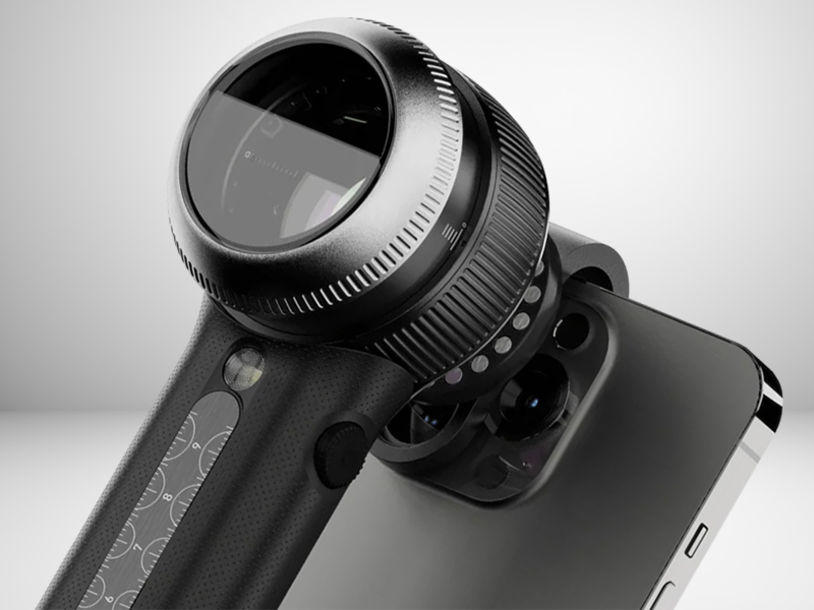
Dermoscopy lets a doctor examine skin structures that are not visible to the naked eye. A dermoscopic photograph records that close-up view so it can be reviewed and compared with later images.
When dermoscopic photography is used
For patients with many moles or higher melanoma risk, dermoscopic photography may be used as part of a broader monitoring strategy such as mole mapping, where images of the skin are compared over time.
We take dermoscopic photos of atypical skin lesions: suspicious, unusual-looking or “ugly duckling” spots. These spots don’t look like typical skin cancers, but we must consider the possibility that they are.
Some patients also monitor their moles between appointments using personal photographs of moles. While these images cannot replace dermoscopic examination, carefully taken photographs can help people notice new or changing lesions that should be assessed during a professional skin check.
How dermoscopic photography helps assess a skin lesion
During your consultation your doctor examines suspicious spots using dermoscopy. If needed, a dermoscopic photograph may be taken to document the lesion and allow more detailed evaluation.
A high-magnification dermoscopic photograph allows:
- The doctor to review the lesion carefully during the consultation.
- Comparison with future photographs to detect subtle changes over time.
- Computer analysis using an AI algorithm, which can provide a supplementary “second opinion” in certain situations (Esteva 2017).
Sequential dermoscopic photography
Your doctor will recommend whether a lesion needs a biopsy or can be monitored. If monitoring is appropriate, they will explain when to return; the interval depends on the lesion and your circumstances.
Comparing images can reveal subtle changes and help avoid unnecessary removal of unchanged benign lesions. A change may lead to a biopsy, but photographs alone cannot rule out skin cancer. Have a new concern assessed rather than waiting for a scheduled photograph.
Accessing your dermoscopic photographs
Your photos are stored in the DermEngine/MoleScope system, which means you have access to them. Your own photographs may help you notice changes, but they do not replace clinical assessment.
If you have your skin checked at another skin clinic in future, you can provide your photos to your new clinic, to give them the opportunity to diagnose skin cancers.
Warnings
Dermoscopic photography significantly increases the ability of the doctor to diagnose early melanoma. However, it is still possible for melanoma and other skin cancers to be missed even with dermoscopic photography repeated at the recommended interval: a low-risk appearance on dermoscopic photography does not always rule out skin cancer. The best way of diagnosing skin cancer is a skin biopsy, which should be performed if there is a reasonable suspicion of skin cancer.
References
Esteva A, Kuprel B, Novoa R, Ko J et al Dermatologist-level classification of skin cancer with deep neural networks. Nature volume 542, pages 115–118. 2017
Salerni G, Terán T, Alonso C, Fernández-Bussy R The role of dermoscopy and digital dermoscopy in the clinical diagnosis of melanoma: clinical and dermoscopic features of 99 consecutive primary melanomas. Dermatology Practical and Conceptual 2014
Rademaker M, Oakley A Digital monitoring by whole body photography and sequential digital dermoscopy detects thinner melanomas. Journal of Primary Health Care 2(4) 268 – 272, 2010.
Menzies S, Guitera P, Soyer P et al What is the role of sequential digital dermoscopy imaging in melanoma diagnosis? Clinical practice guidelines for the diagnosis and management of melanoma, 2018.

MBBS, MA (Virtual Comm), Grad Cert Hlth Info, Grad Dip Comp Inf Sci
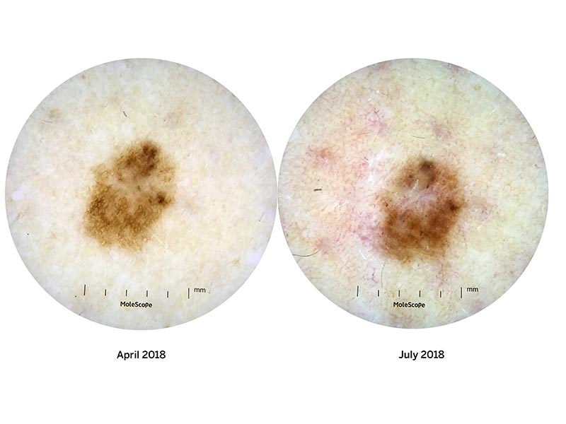
This lesion has changed on sequential dermoscopic photography over a period of 3 months. It was biopsied and discovered to be a melanoma in situ (i.e. diagnosed early and cured)
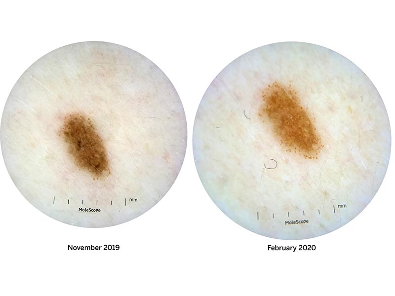
This lesion grew significantly over a period of three months. Biopsy showed that it was a melanoma in situ.
Concerned about a suspicious mole or changing spot?
Dermoscopic examination allows doctors to detect subtle features of skin cancer that are not visible to the naked eye. If you have a new or changing lesion, a professional skin check can assess it using dermoscopy and, if necessary, dermoscopic photography.
Find out about a spot assessment